Oral Histopathology
Microscopic examination of oral tissue samples to support accurate diagnosis of lesions, abnormalities and other oral pathological conditions.
What oral histopathology involves
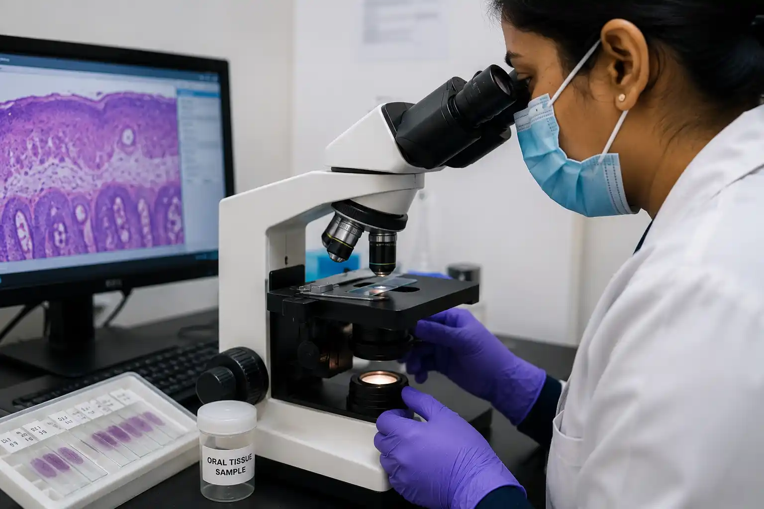
When your dentist notices a lesion, growth or abnormal tissue in the mouth, a small sample is taken and examined under a microscope. This examination looks at the tissue's cellular structure to help identify the nature of the condition and guide the right course of treatment.
Common uses: evaluating oral lesions, ulcers or growths, investigating abnormal tissue changes, and confirming a diagnosis before or after a surgical procedure.
Is oral histopathology right for your patient?
Persistent lesions
An oral lesion, ulcer or growth that hasn't resolved and needs investigation to understand its nature.
Post-surgical confirmation
Confirming the nature of tissue removed during an oral surgical procedure.
Unusual tissue changes
Any abnormal change in colour, texture or growth pattern in the oral tissues that warrants a closer look.
Sample collection
Your dentist takes a small tissue sample from the area of concern and sends it for laboratory examination.
Microscopic analysis
The tissue is examined under a microscope to assess its cellular structure and identify the condition.
Report
Findings are compiled into a diagnostic report shared with your referring dentist.
Coordination
Works alongside your dentist's clinical examination as part of a complete diagnostic picture.