OPG vs CBCT: What's the Difference?
By Dr. 2, [Full Name], [Qualification] · Reviewed 12 August 2026
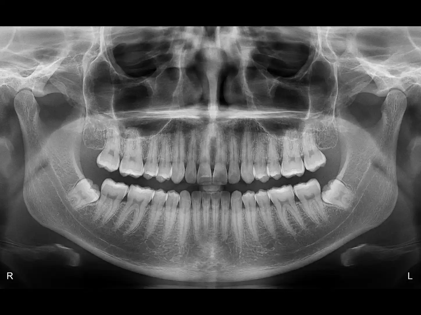
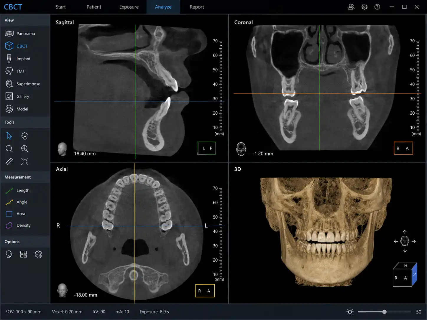
An OPG is a single flat panoramic X-ray showing both jaws in one 2D image, best for routine screening — a CBCT scan produces a full 3D reconstruction of bone, nerves and roots, used when a dentist needs precise depth and volume information, such as before placing an implant.
What an OPG shows
An orthopantomogram (OPG) captures your entire jaw — both arches, all teeth, the sinuses and the TMJ — in a single rotating exposure. It's fast, low-dose, and gives your dentist a broad overview: cavities, bone loss, impacted teeth, cysts, or anything unusual that warrants a closer look.
What a CBCT shows
Cone Beam CT (CBCT) takes hundreds of individual images and reconstructs them into a 3D model. Instead of one flat picture, your dentist can scroll through cross-sections at any angle — seeing exactly how much bone surrounds a potential implant site, precisely where a nerve canal runs, or whether a root canal has a hidden extra canal that a flat X-ray would miss.
How dentists choose between them
OPG is almost always the first scan — a broad, low-dose screening tool used at routine check-ups, before orthodontic treatment, or to get a first look at wisdom teeth. CBCT is reserved for cases where 3D precision actually changes the treatment plan: implant placement, complex root canal retreatment, impacted tooth surgery, or TMJ and airway evaluation. Many patients have an OPG first, and a CBCT is ordered only if that initial image raises a question that needs 3D detail to answer.
Radiation dose comparison
Both are low-dose compared to a medical CT scan. OPG has one of the lowest doses in dental imaging. CBCT delivers a higher dose than OPG, but our units are calibrated to ALARA protocols with the field of view sized to the specific clinical question, keeping exposure to the minimum needed for a diagnostic-quality image.
Related: Learn about OPG · Learn about 3D CBCT · Read our full FAQ